Figure 6 from Femoral Hernia: A Review of the Clinical Anatomy and

By A Mystery Man Writer
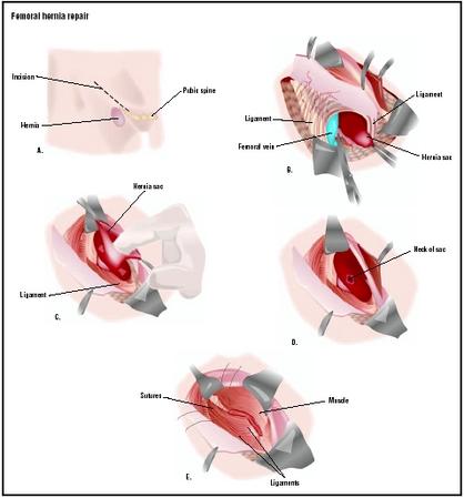
Figure 6. Femoral hernia repair in clean operation. (a) The narrow side of the mesh is sutured to Cooper’s ligament; (b) The mesh is sutured to the iliopubic tract or shelving portion of the inguinal ligament; (c) The posterior wall of the inguinal canal is reinforced, as in Lichtenstein’s repair. - "Femoral Hernia: A Review of the Clinical Anatomy and Surgical Treatment"
Hernia - Physiopedia

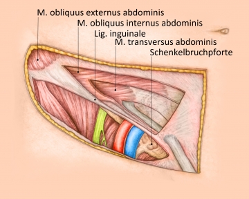
Figure 4 from Femoral Hernia: A Review of the Clinical Anatomy and Surgical Treatment

Embryonic developmental process and clinical anatomy of the preperitoneal fascia and its clinical significance

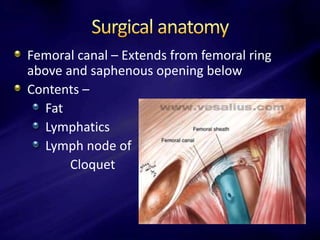
Femoral Hernia - A Review of Clinical Anatomy
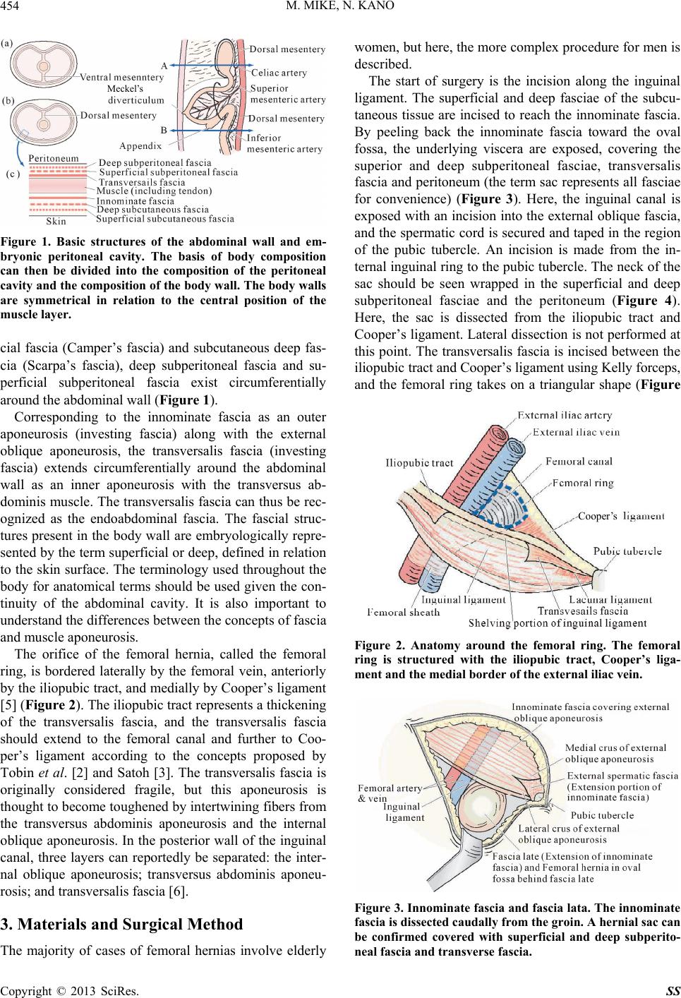
Femoral Hernia: A Review of the Clinical Anatomy and Surgical Treatment

Schematic view of right femoral region illustrating variants of femoral
Abdominal Hernia - Epigastric - Spigelian - Obturator - TeachMeSurgery
Femoral Hernia - A Review of Clinical Anatomy
Femoral hernia

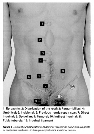
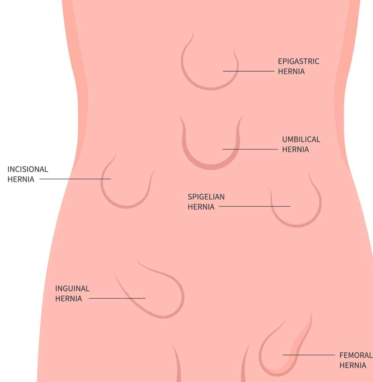
The anatomical locations of the groin hernia defects. 1: Lateral

Figure 12 from Femoral hernia repair.
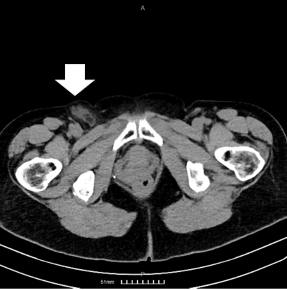
Cureus, Femoral Hernia Containing a Strangulated Appendix: A Hybrid Approach
- Anatomy - Femoral hernia repair – TIPP technique
- PDF] Tension free femoral hernia repair with plug

- Femoral Hernia Repair - procedure, recovery, blood, pain, complications, adults, time, infection
- Repair of Inguinal and Femoral Hernias
- Incarcerated Femoral Hernia and the Ovary Found Within It - SAGES Abstract Archives