PLAQUE-TYPE BLUE NEVUS – A SINGULAR VARIANT OF BLUE NEVUS

By A Mystery Man Writer
A 48 year-old male who had a blue-gray plaque with multinodularity arising in the left scapular region is presented, showing the presence of a blue nevus predominantly of the common type, and focal cellular areas in its deeper portion. Introduction: Plaque-type blue nevus is a rare variant of blue nevus, presenting usually as blue-gray plaque with superimposed nodules, beginning at birth or in early childhood and becoming stable during puberty. Its clinical, dermoscopic and even histopathological features may be worrisome and distinction from malignant blue nevus can be difficult, although its prognosis is generally favourable. Case Report: We present a 48 year-old male who had a blue-gray plaque with multinodularity arising in the left scapular region. The lesion had been present since birth, becoming stable during adolescence. Dermoscopy revealed a central structureless polychromatic plaque (blue, gray, black and brown areas) and extensive areas of blue-whitish veil. Adjacent to the central lesion, small blue satellite globules could be seen. Incisional biopsy showed a common blue nevus, and complete excision of the lesion confirmed the presence of a blue nevus predominantly of the common type, and focal cellular areas in its deeper portion. The patient is so far free of recurrence. Conclusion: There are many variants of blue nevus, and plaque-type blue nevus is one of the rarest. Classically regarded as having good prognosis, recent case-reports have shown a tendency towards local and lymph-node recurrence, the reason why recognition of this entity and appropriate follow-up are important.

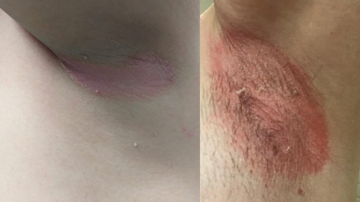
EBN (blue arrows) on the left shoulder in a zosteriform distribution.

Blue Nevus - Academic Dermatology of Nevada

Compound blue nevus”: A reappraisal of “superficial blue nevus with prominent intraepidermal dendritic melanocytes” with emphasis on dermoscopic and histopathologic features - ScienceDirect

Surgical Treatment of Nevi and Melanoma in the Pediatric Age
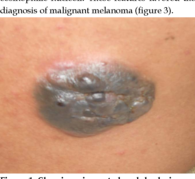
Blue Melanoma Simulating Blue Nevus

Agminated Blue Nevus Arising in a Nevus Spilus

SF3B1 and BAP1 mutations in blue nevus-like melanoma - ScienceDirect
Frontiers The Spectrum of Spitz Melanocytic Lesions: From Morphologic Diagnosis to Molecular Classification

DermDx: Pigmented Plaque on the Upper Back - Clinical Advisor

PDF) Beware of the “Blue Nevus”: A Case Report

SciELO - Brasil - Atypical cellular blue nevus or malignant blue nevus?* Atypical cellular blue nevus or malignant blue nevus?*

Histology revealing a blue nevus.