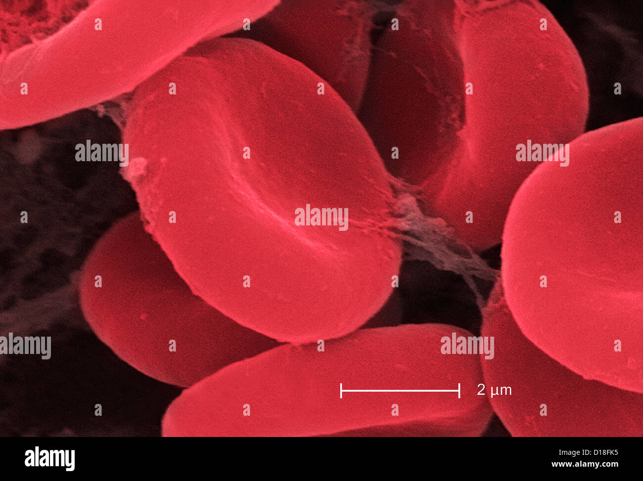
This scanning electron micrograph (SEM) depicted a number of red

By A Mystery Man Writer
Download this stock image: This scanning electron micrograph (SEM) depicted a number of red blood cells found enmeshed in a fibrinous matrix on the luminal surface of an indwelling vascular catheter; Magnified 11432x Note the biconcave cytomorphologic shape of each erythrocyte, which increases the surface area of these hemoglobin-filled cells, thereby, promoting a greater degree of gas exchange, which is their primary function in an in vivo setting. In their adult phase, these cells possess no nucleus. What appears to be irregularly-shaped chunks of debris, are actually fibrin clumps, which when inside the living organi - 2BE0H0B from Alamy's library of millions of high resolution stock photos, illustrations and vectors.
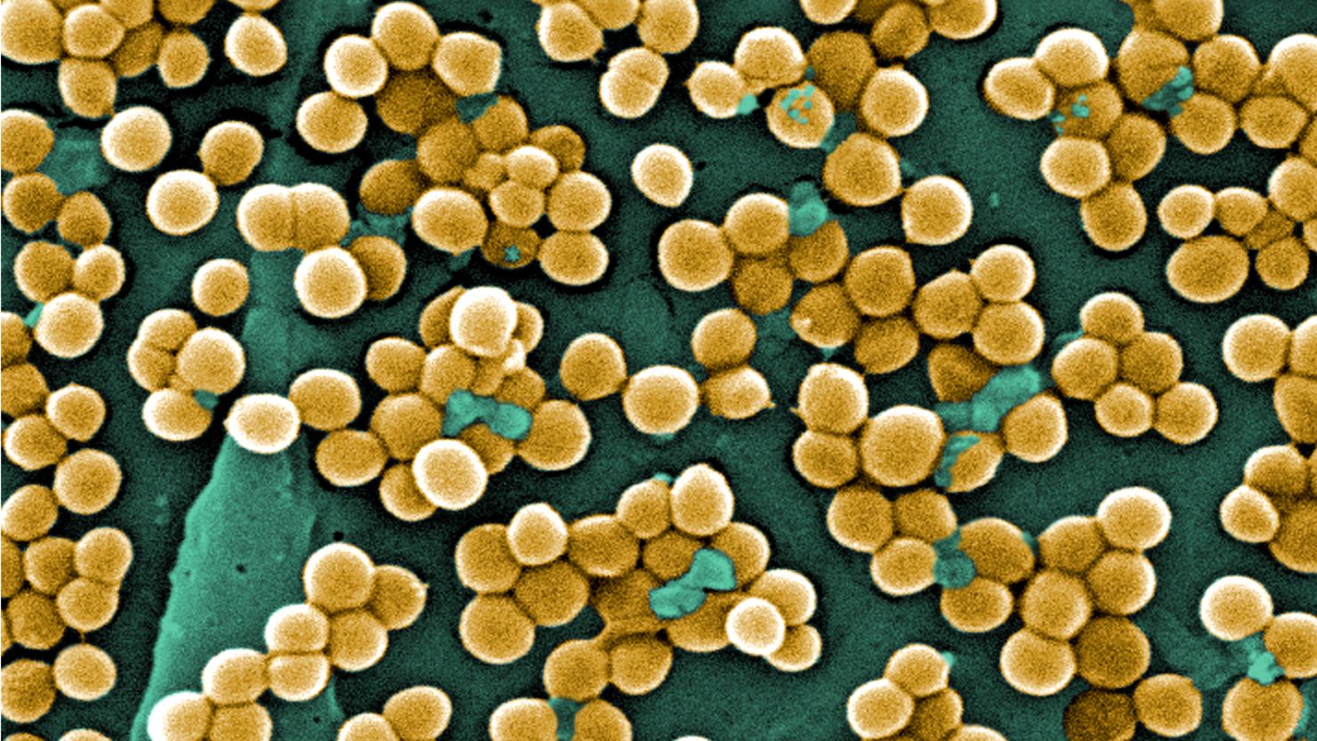
MRSA Fast Facts

35 Indwelling Catheter Photos & High Res Pictures - Getty Images

Microscopic silk present on the exoskeleton cuticle of the honey bee, SEM, Stock Photo, Picture And Rights Managed Image. Pic. BSI-0120006

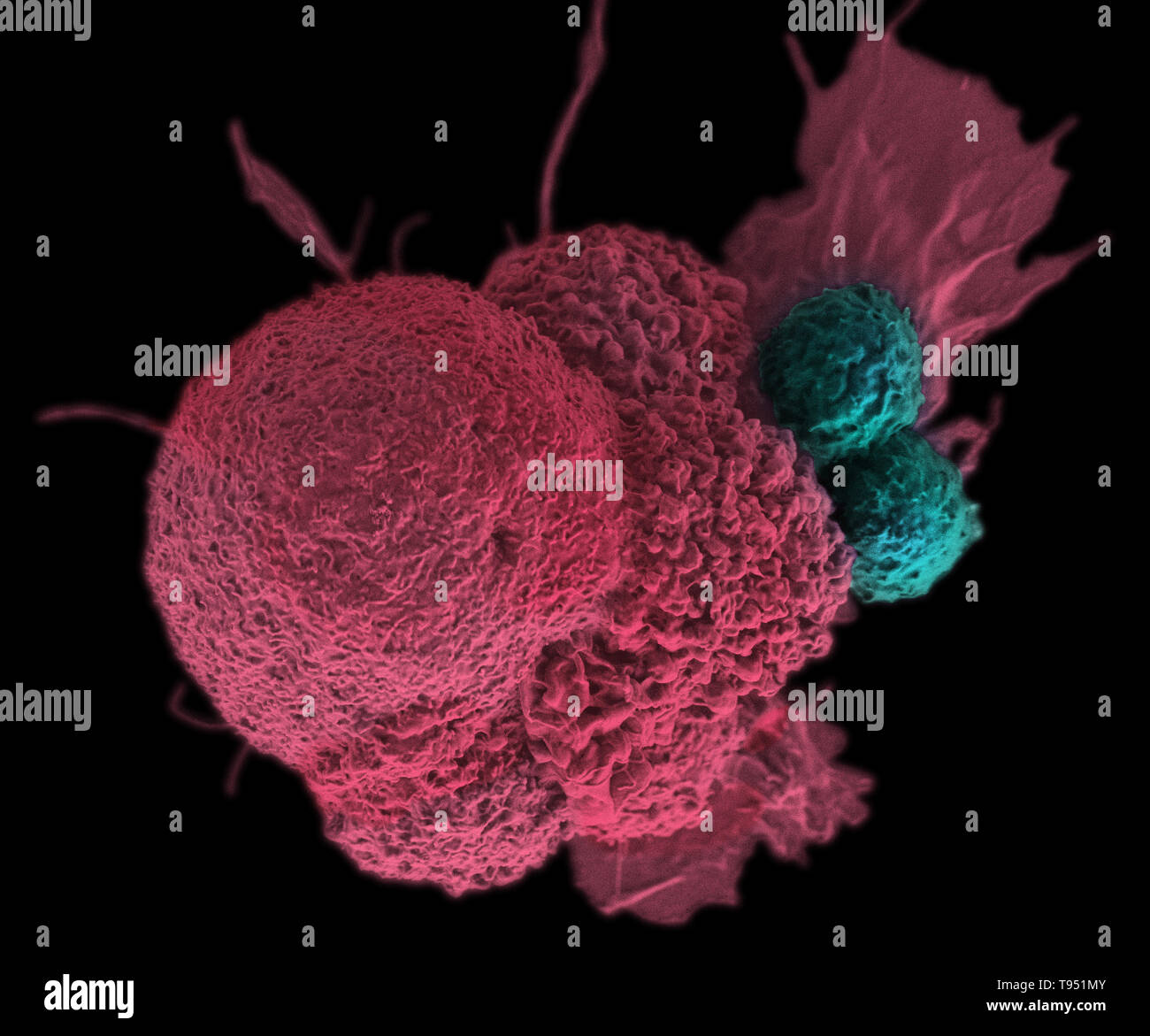
Colorized sem horizontal hi-res stock photography and images - Alamy
Sem blood hi-res stock photography and images - Alamy
Electron micrograph hi-res stock photography and images - Alamy
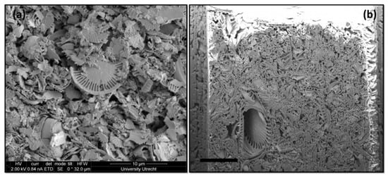
Minerals, Free Full-Text

Sem blood hi-res stock photography and images - Alamy

Public Domain Picture, This scanning electron micrograph (SEM) depicted a number of red blood cells found enmeshed in a fibrinous matrix on the luminal surface of, ID: 13518540025049

A. Scanning electron microscope picture of red blood cells subjected
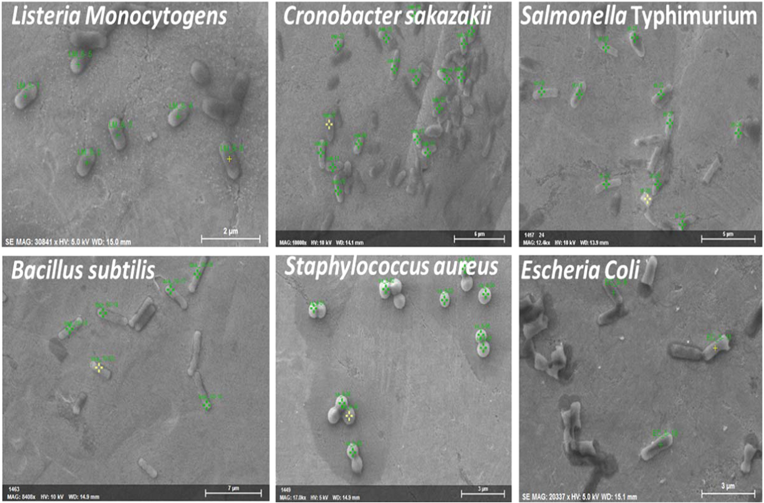
Power of Scanning Electron Microscopy and Energy Dispersive X-Ray Analysis in Rapid Microbial Detection and Identification at the Single Cell Level
- HEMO Shapewear Women's Tummy Control Slimming Panties Hourglass

- HEMO Shapewear Women's Tummy Control Shapewear Light Satin Briefs

- HEMO Body Mujer Shapewear Sin Tirantes Sin Espalda con Un Traje

- HEMO Body Saper Bodysuit Shapewear Light Satin Panties Briefs High

- Effects of CO poisoning on hemoglobin-oxygen dissociation curve. In

- Erika Villavicencio - Production Manager and Product Developer
- cotton organdy chocolate pintuck-pink
- Frenchtrendz Buy Frenchtrendz Cotton Spandex Fuchsia Capri Online

- TriDri Lightweight Training Leggings TR017-Sports Gym Running Jogging Leggings

- Saks Fifth Avenue in NYC Just Unveiled Its First-Ever Kids' Floor