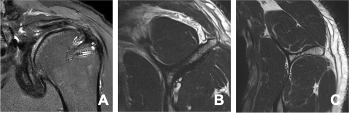
Modes of rotator cuff failure. Notes: ( A ) Intact repair on MRI. Note

By A Mystery Man Writer
Download scientific diagram | Modes of rotator cuff failure. Notes: ( A ) Intact repair on MRI. Note the position of the musculotendinous (MT) junction at the midpoint of the humeral head. ( B ) Failure with defect on MRI. Note the defect at the greater tuberosity and the significantly retracted position of the MT junction. ( C ) Failure with continuity on MRI. Note the continuous tissue extending to the greater tuberosity despite significant retraction of the MT junction. White arrows in A–C show position of the MT junction. ( D ) Illustration of failure with defect. ( E ) Illustration of failure with continuity. Illustrations by David Schumick, BS, CMI. Reprinted with the permission of the Cleveland Clinic Center for Medical Art and Photography © 2015. All Rights Reserved. Abbreviation: MRI, magnetic resonance imaging. from publication: Rotator cuff repair: Challenges and solutions | Each year, 250,000 rotator cuff repairs are performed in the United States at a cost of $3 billion. Despite advancements in repair techniques and rehabilitation, 20%-70% of repairs continue to undergo structural failure; however, there is a poor correlation between clinical | Rotator Cuff, Repair and Platelet Rich Plasma | ResearchGate, the professional network for scientists.
Rotator cuff tear patterns: MRI appearance and its surgical

Nontendinous healing after repairing of retracted rotator cuff

Can Preoperative Magnetic Resonance Imaging Predict the
Frontiers Decellularized biological matrices for the repair of rotator cuff lesions: a systematic review of preclinical in vivo studies

Modes of rotator cuff failure. Notes: ( A ) Intact repair on MRI

Michael AMINI, Cleveland Clinic, OH, Department of Orthopaedic Surgery

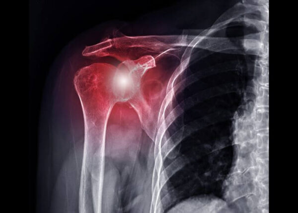
Rotator cuff injury
Rotator cuff tear patterns: MRI appearance and its surgical
Rotator cuff tear patterns: MRI appearance and its surgical
Intact revision rotator cuff repair stabilizes muscle atrophy and

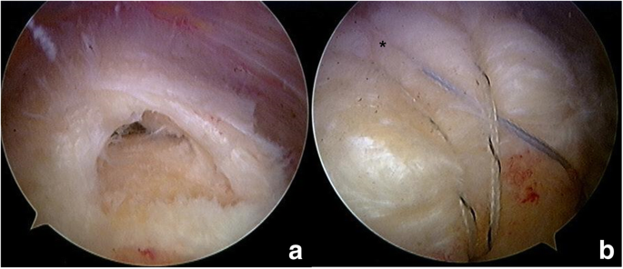
Studies of commercially available scaffolds in interposition of

Arce 2013, PDF, Shoulder
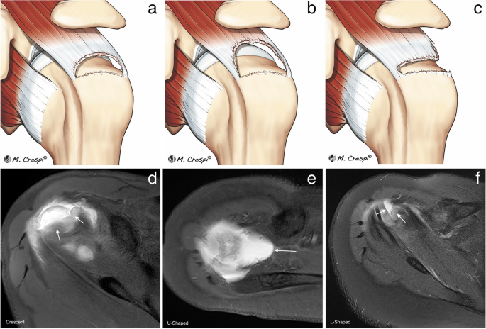
Rotator cuff tear patterns: MRI appearance and its surgical
- Delaying rotator cuff repair surgery more than 12 months may be
- JCM, Free Full-Text
- Revision Rotator Cuff Repair, Shoulder Surgeon
- Clinical outcomes and repair integrity of arthroscopic rotator cuff repair using suture-bridge technique with or without medial tying: prospective comparative study, Journal of Orthopaedic Surgery and Research
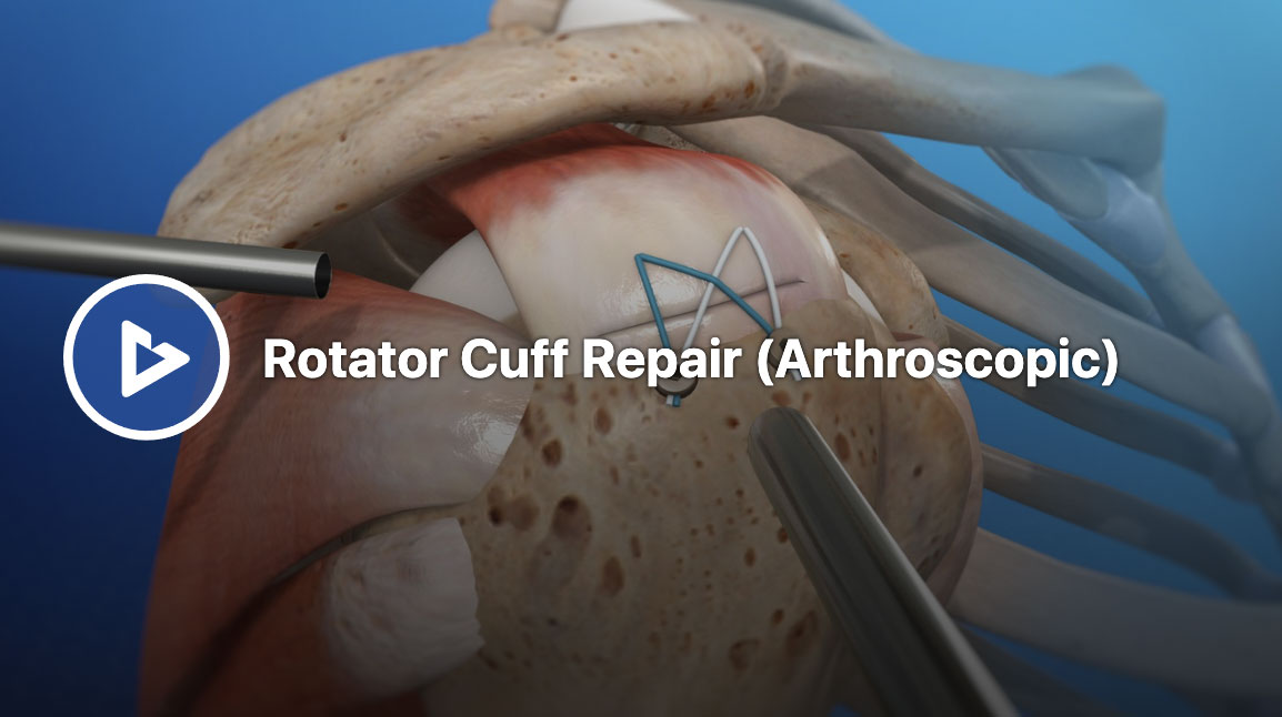
- Arthroscopic Rotator Cuff Repair
- Fashion Women's Plus Size Tummy Control Panties Thigh Slimmer
/product/17/167865/1.jpg?9926)
- Crystal Lace Necklace Patterns, Bead Weaving Technique

- Natori Women's Feathers Refresh Full-Fit Underwire Bra 734331

- Bras for Women Buckle Middle Aged and Elderly Underwear Women Smooth No Underwire Wide Strap Vest Type New (A, 36) at Women's Clothing store

- I Love Vivier Pumps in Suede Black Woman RVW53024540O2041B999
