Histology, microscopy, anatomy and disease: Week 3: 2.1
By A Mystery Man Writer
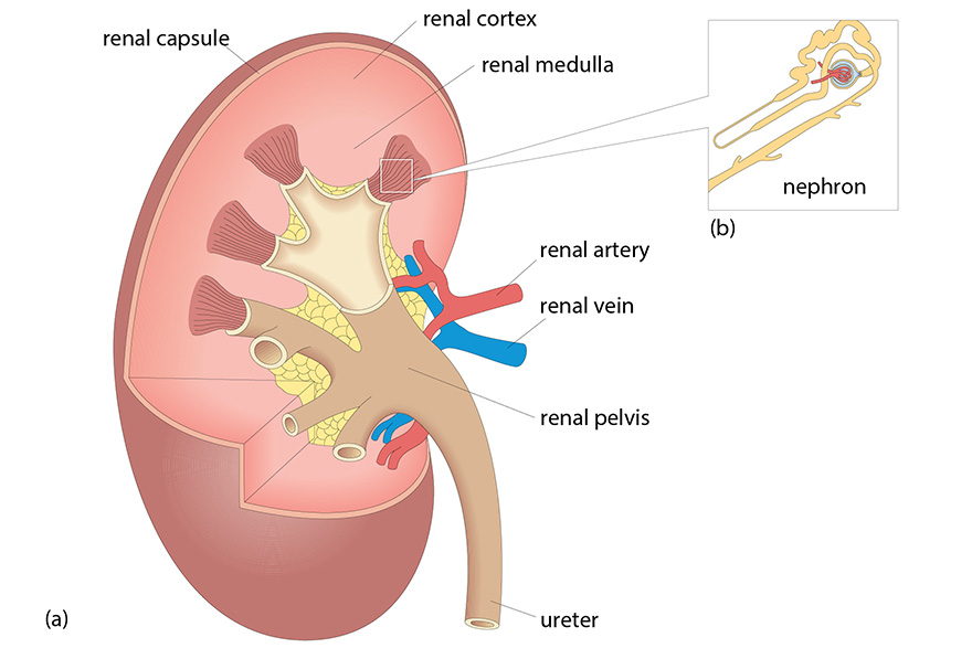
Histology, microscopy, anatomy and disease: Week 3: Figure 10 Diagram of the kidney structure with an enlarged diagram of a nephron. The ureter carries urine from the kidney to the bladder.
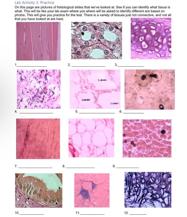
Solved Lab Activity 3: Practice On this page are pictures of
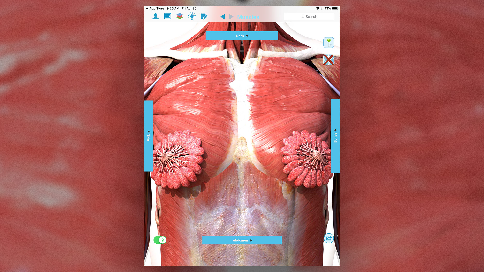
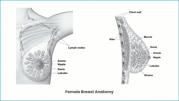
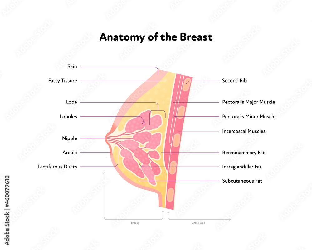
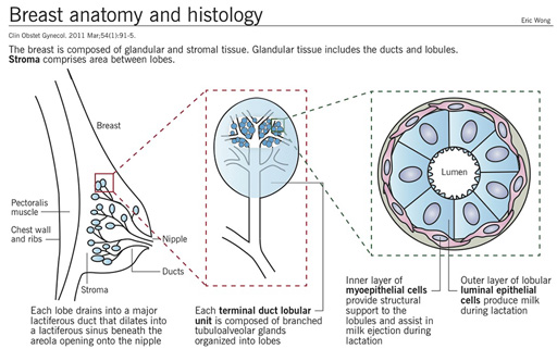
High quality, high discounts Breast Anatomy: Milk Ducts, Tissue

GI Pathology Amboss Q&A Flashcards
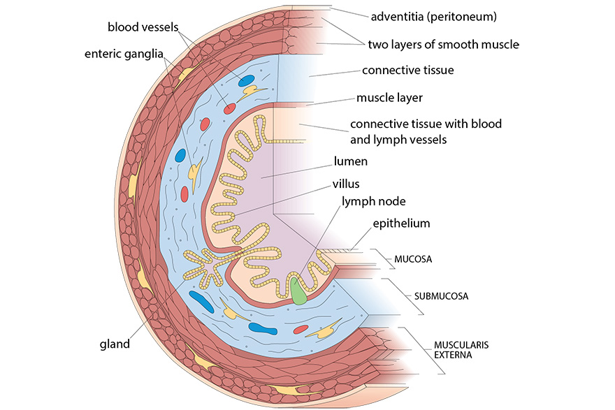
Histology, microscopy, anatomy and disease: Week 2: Figure 3 Cross-section of the gut showing the arrangement of the different tissue elements at the level of the ileum (small intestine).

Infection of 3D Brain Organoids with Human Pathogenic Viruses Under Biosafety Level-3 Conditions with Subsequent Inactivation to Study Viral Replication, Pathomechanisms, and Other Viral Infection-Mediated Effects

2.4.3 Skin Pathology I Flashcards by Priyanka Bhandari
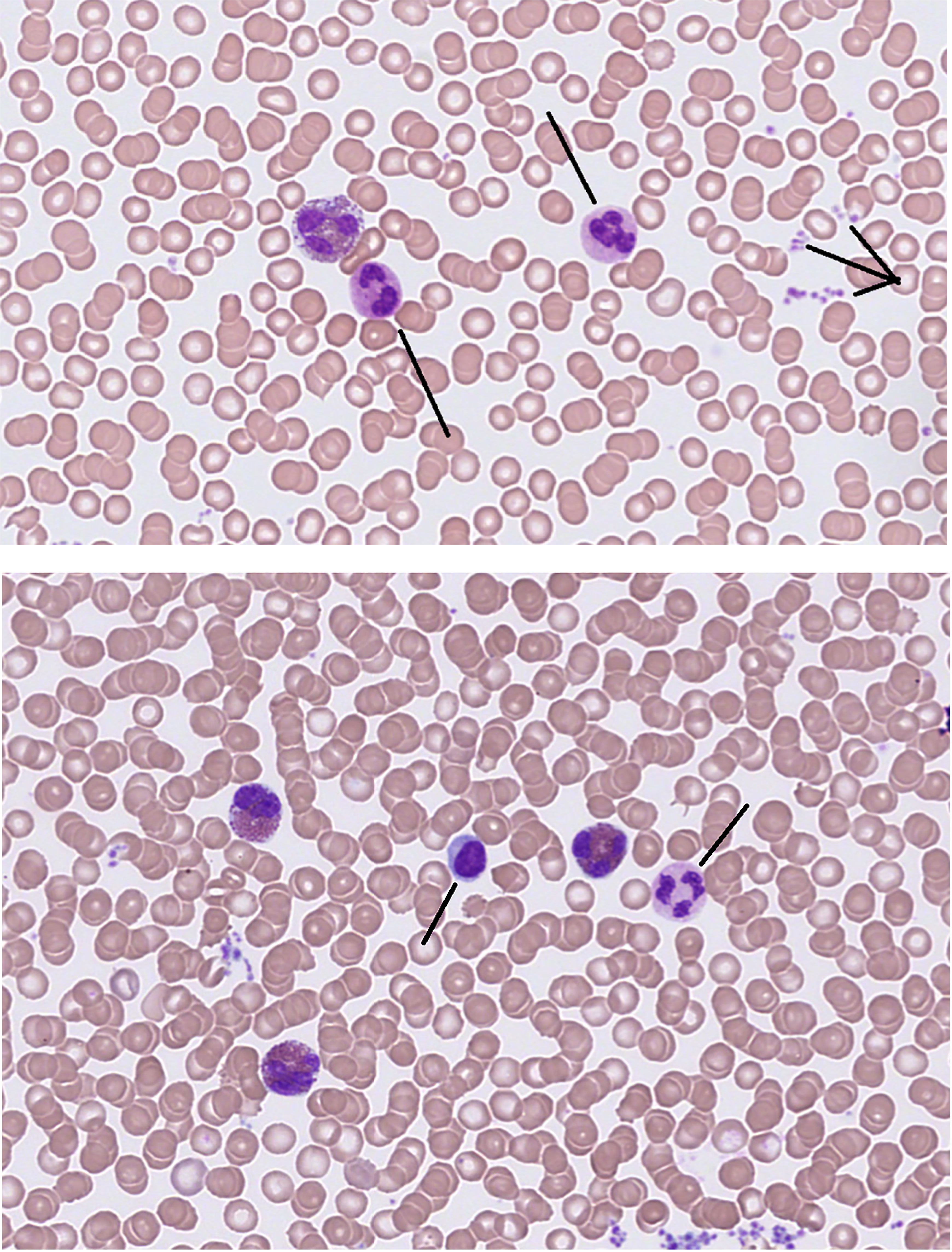
Frontiers The multidisciplinary approach to eosinophilia

Anatomy, Histology, and Normal Imaging of the Endometrium
Human Pathology Nikon's MicroscopyU
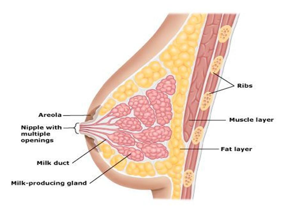
High quality, high discounts Breast Anatomy: Milk Ducts, Tissue

High quality, high discounts Breast Anatomy: Milk Ducts, Tissue

Toxins, Free Full-Text
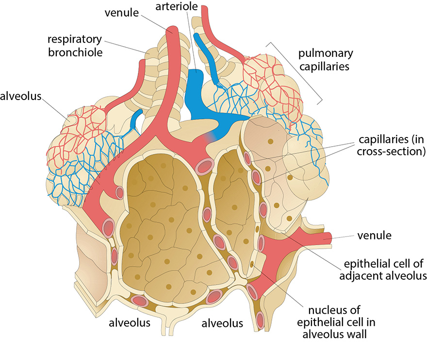
Histology, microscopy, anatomy and disease: Week 3: Figure 2 Schematic diagram of an alveolus, in contact with pulmonary capillaries (Villee, 1989).