Coronal and axial slices displaying the IFG area that showed
By A Mystery Man Writer
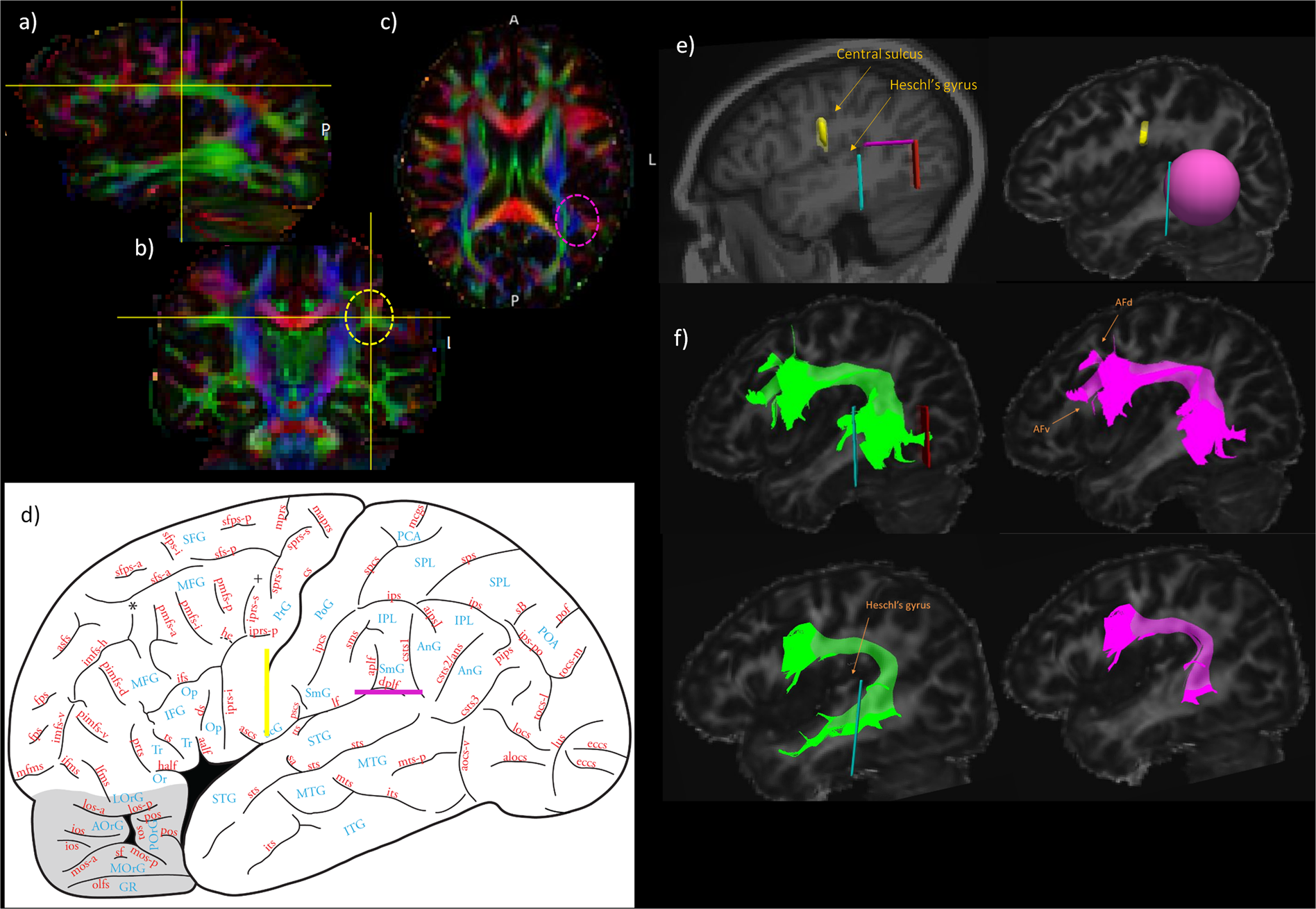
Dissociating the white matter tracts connecting the temporo-parietal cortical region with frontal cortex using diffusion tractography
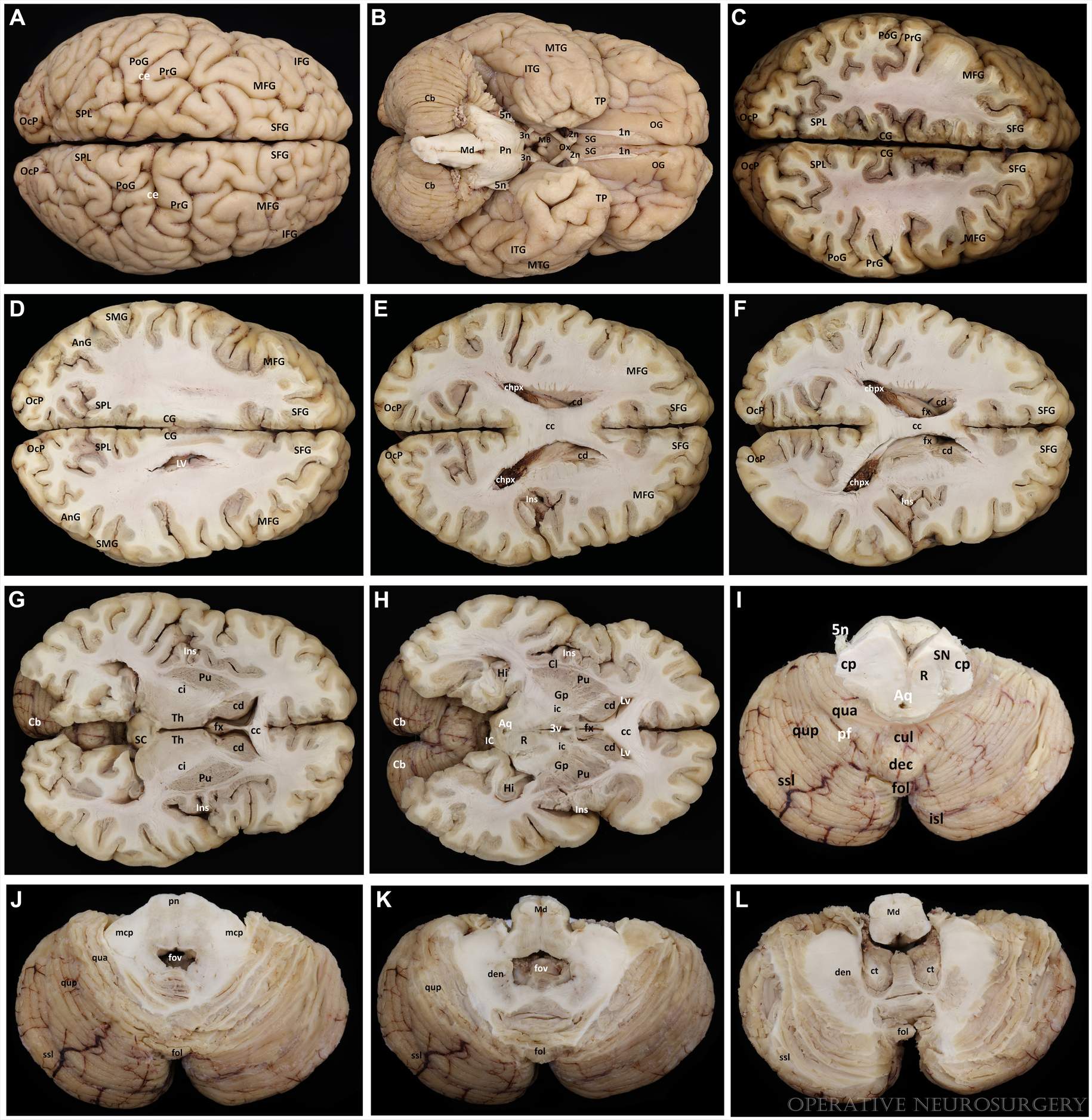
3D Modeling and Extended Reality Simulations of the Cross-sectional Anatomy of the Cerebrum, Cerebellum, and Brainstem

Sagittal, coronal, and/or axial slices across the main areas of
David ZALD, Professor (Full), Ph.D.

Full article: Functional Magnetic Resonance Imaging at 3T as a Clinical Tool in Patients with Intracranial Tumors
Relationships between Eye Movements during Sentence Reading Comprehension, Word Spelling and Reading, and DTI and fmri Connectivity In Students with and without Dysgraphia or Dyslexia

José PARDO Professor (Full); Director, Cognitive Neuroimaging

Neural correlates of recovery from aphasia after damage to left inferior frontal cortex

Reading musical notation versus English letters: Mapping brain activation with MEG - Ching-I Lu, Margaret L. Greenwald, Yung-Yang Lin, Susan M. Bowyer, 2019
- Order IFG Vision Bra, White Online at Special Price in Pakistan

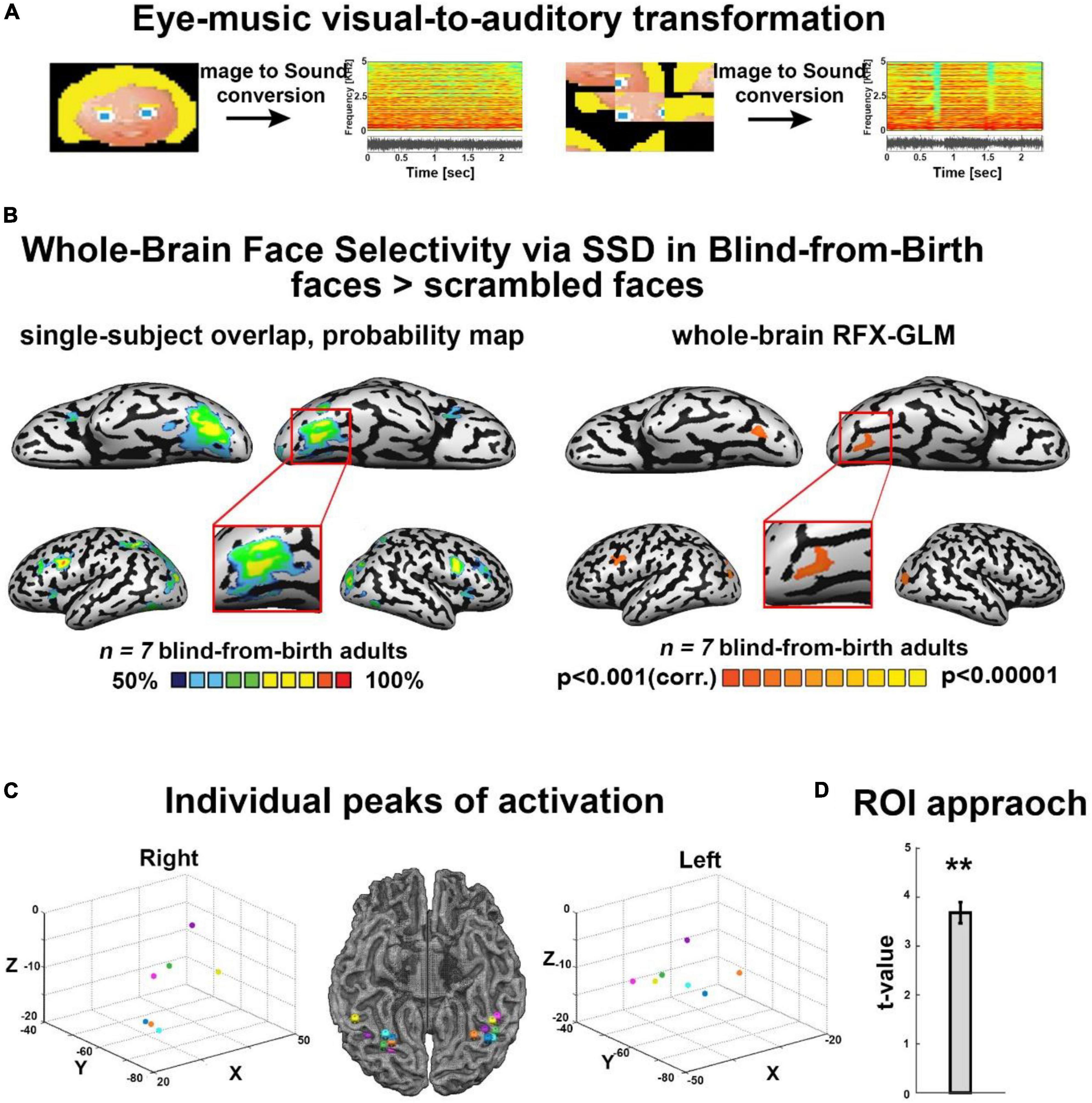
- Frontiers Face shape processing via visual-to-auditory sensory substitution activates regions within the face processing networks in the absence of visual experience
- CRONY BEIGE SHORT BRA Slimming body shaper and Beauty Inner wear
- IFG Companies Benefits

- IFG - The iFish Group - Greater Sacramento Economic Council