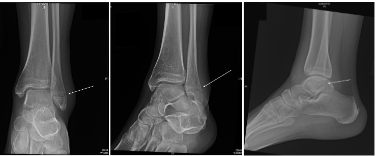
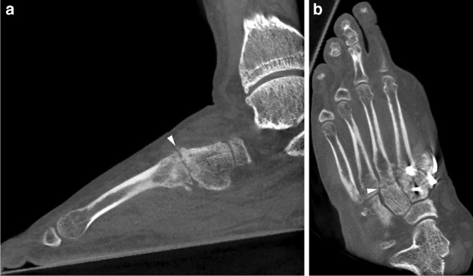
Right calcaneal fracture in a 36-year-old male patient treated with

By A Mystery Man Writer
Download scientific diagram | Right calcaneal fracture in a 36-year-old male patient treated with sinus tarsi approach. a, b Preoperative X-ray films. c, d Preoperative CT. e Marking for surgical incision. f No. 1 K-wire was drilled transversely into the posterior part of the calcaneus, traction of the K-wire was directed in a backward, downward and valgus motion, aiming to restore the length, height and Bohler’s Angle of the calcaneus. g No. 2 K-wire, No. 3 K-wire and No.4 K-wire were used to restore and fix the collapsing articular facet of subtalar joint. h The plate would be placed. i Intraoperative fluoroscopy confirming the position of the plate and screws. j Postoperative incision condition, No. 1 and No. 2 auxiliary incisions were used to help place the screws. k, l X-ray films 1 day after operation. m CT one day after operation. n, o X-ray films 6 months after operation from publication: Same wound complications between extensile lateral approach and sinus tarsi approach for displaced intra-articular calcaneal fractures with the same locking compression plates fixation: a 9-year follow-up of 384 patients | Purpose Some previous studies have demonstrated that the sinus tarsi approach (STA) is a better therapeutic method than the extensile lateral approach (ELA) for displaced intra-articular calcaneal fractures. In a number of those previous studies, two different implants were | Heel, Wounds and Displacement (Psychology) | ResearchGate, the professional network for scientists.

A 36-year-old male patient had a right calcaneal fracture as a
Lateral Malleolar Fracture Published in Orthopedic Reviews
Clinical Research on Foot & Ankle - Intra-Articular Calcaneal

10000 PDFs Review articles in STA
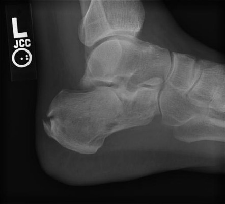
Calcaneal fracture, Radiology Reference Article

10000 PDFs Review articles in STA

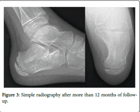
Bony destructive injuries of the calcaneus: long-term results of a

Comparing less invasive plate fixation versus screw fixation of

Linbo Zhuang's research works First Affiliated Hospital of China Medical University, Shenyang and other places

Calcaneus Fractures - Trauma - Orthobullets

A) Lateral X-ray of calcaneus, obvious displacement of calcaneal
Imaging of osteoarthritis from the ankle through the midfoot

Calcaneus Fractures - Trauma - Orthobullets
- Dia Quente De Verão. Termômetro De Celsius E Fahrenheit Na Areia. Temperatura Ambiente Mais 36 Foto de Stock - Imagem de célsio, cadeira: 274536342

- Zelotes F-36 vertical sem fio 2.4g bluetooth mouse cor cheia luz 8 chave de programação 2400dpi jogo mouse 730mah bateria de lítio - AliExpress

- Disco De Lixa Fibra Metalite F212 180mm Grão 36 C/ 10 Pçs Norton - Disco de Lixa / Flap - Magazine Luiza
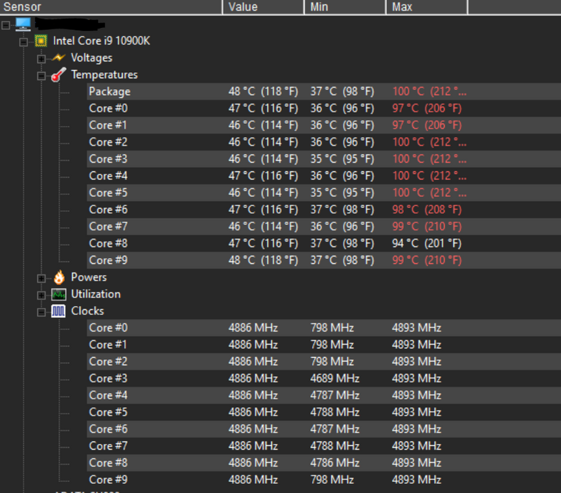
- Omen 30L CPU get's burning hot and sounds like a starting 74 - HP Support Community - 7896691
- Could Your Car Become a Heat Trap? - FOUR PAWS in US - Global

- Sexy Honey Moon Bridal Wedding Short Net Nighty - R162 - Online Shopping in Pakistan - Online Shopping in Pakistan - NIGHTYnight

- Man Writing The Word Assurance Stock Photo, Picture and Royalty

- Sonari Mothercare Women Maternity/Nursing Non Padded Bra - Buy

- mama agnes short movie

- Skull & Bones indigo bandana brief – Haut Underwear