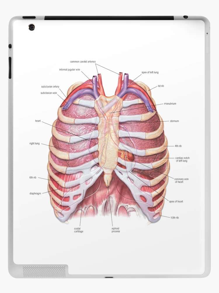
Figure 3 from Relevant surgical anatomy of the chest wall.

By A Mystery Man Writer
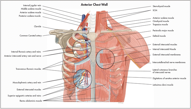
Fig. 3. Anterior chest wall showing the sternum. Note where the costal cartilages articulate with the sternum. In the intercostal space lie different structures: several kinds of intercostal muscles, intercostal arteries and associated veins, lymphatics, and nerves. (From Rendina EA, Ciccone AM. The intercostal space. Thorac Surg Clin 2007;17(4):491e501; with permission.) - "Relevant surgical anatomy of the chest wall."

Reasons Why You Would Be Referred To A Thoracic Surgeon

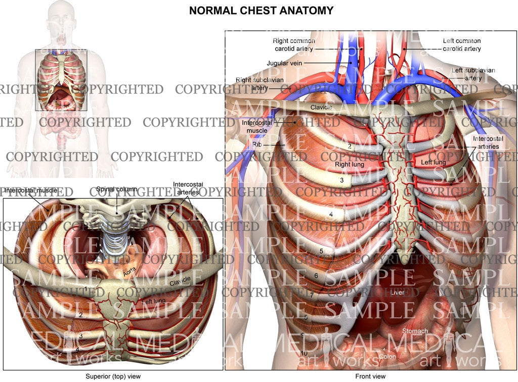
Figure 5 from Relevant surgical anatomy of the chest wall.
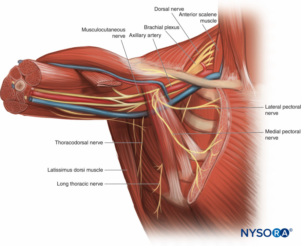
Pectoralis and Serratus Plane Nerve Blocks - NYSORA
Thorax Basicmedical Key
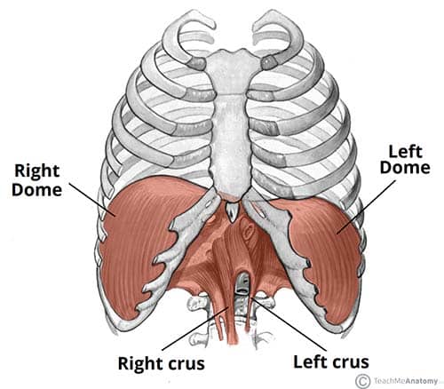
The Diaphragm - Actions - Innervation - TeachMeAnatomy

Figure 3 from Relevant surgical anatomy of the chest wall.
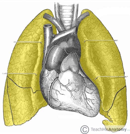
Lung: Anatomy, blood supply, innervation, functions
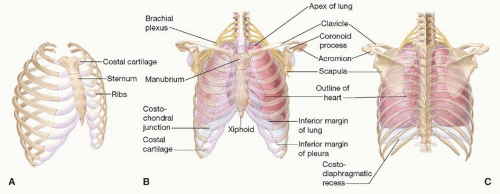
PERTINENT SURGICAL ANATOMY OF THE THORAX AND MEDIASTINUM

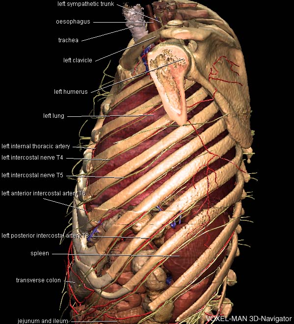
Figure 7 from Relevant surgical anatomy of the chest wall.
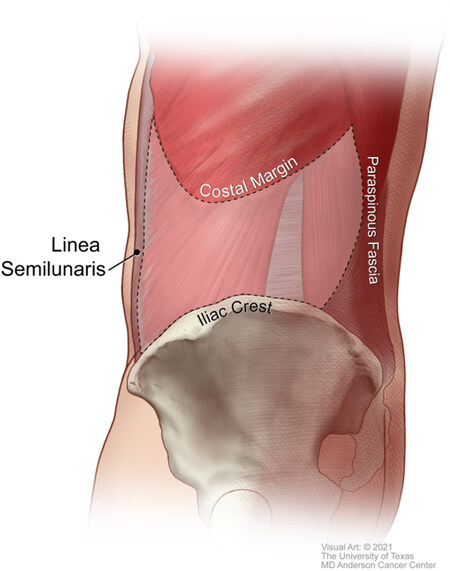
Lateral abdominal wall reconstruction

Introduction to chest wall reconstruction: anatomy and physiology of the chest and indications for chest wall reconstruction. - Abstract - Europe PMC
Chest Wall Reconstruction

Thorax Deformity - an overview
/wp-content/uploads/Anatomical
Surgical Anatomy of the Chest Wall